Chrysomya bezziana - the Old World screwworm fly
Description
The larvae of Old World Screwworm fly (OWSF) Chrysomya bezziana and New World screwworm fly (NWSF) Cochliomyia hominivorax, can be easily confused with each other and with the larvae of other agents of myiasis. Accurate diagnosis involves the identification of larvae extracted from the deepest part of an infested wound. Larvae from the periphery of the wound could be of a secondary myiasis causing species. The mature, third instar larvae are most reliable for morphological identification. Confirmation of OWSF relies on the recognition of a characteristic combination of spinulation, the number of lobes on the anterior spiracles (usually 4-6), and pigmentation of secondary tracheal trunks.
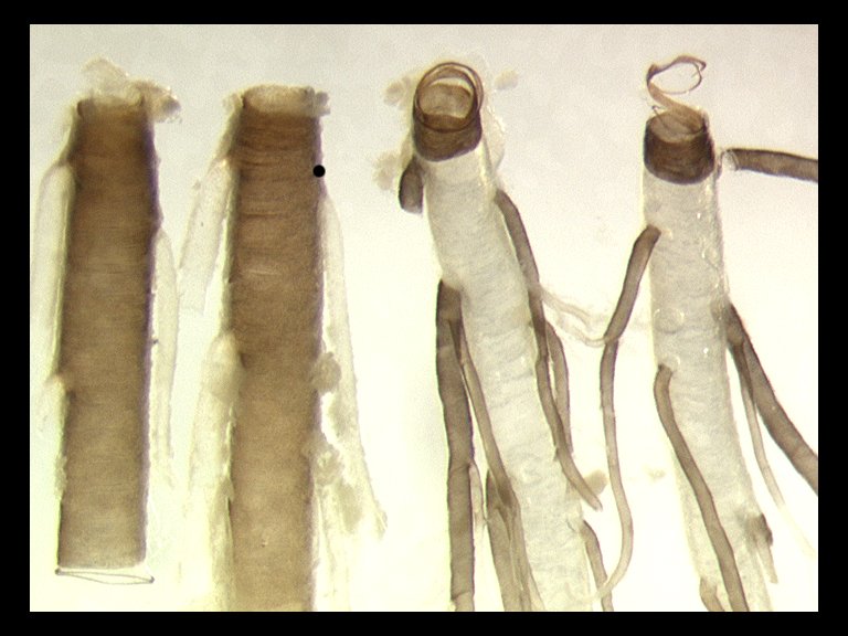
Third instar larvae of OWSF have a robust, typical maggot shape, with a cylindrical body from 6 to17 mm long and from 1.1 to 3.6 mm in diameter, pointed at the anterior end (Spradbery 1991, Laake et al 1936). Fully mature larvae develop a reddish-pink tinge over the creamy white colour of younger larvae. This species has prominent rings of spines around the body and these spines appear large and conspicuous under a microscope when compared with most non-screwworm species, the longest averaging 130 µm. In OWSF the spines always have a single point. The anterior spiracles each have from three to seven branches, but usually from four to six. The latter character should not be used on its own to identify OWSF, because third instar larvae of the obligate myiasis-causing species Wohlfahrtia magnifica (Diptera: Sarcophagidae), whose distribution overlaps that of OWSF in the Middle East, have similarly branched anterior spiracles. Hence, in using any identification key, it is essential that each specimen be taken through the whole key to avoid misidentifications. On the posterior face of the terminal segment of OWSF, the posterior spiracular plates all have a darkly pigmented, incomplete peritreme enclosing three straight, slightly oval-shaped slits, which point towards the break in the peritreme. Of particular diagnostic value in separating OWSF from NWSF are the dorsal tracheal trunks, which extend forwards from the posterior spiracular plates. This feature is seen most easily in living larvae. Those in preservative may need dissection to remove opaque tissues covering the trunks. The dorsal tracheal trunks of OWSF are darkly pigmented only in the twelfth segment. However, in OWSF the secondary trachea branching off the dorsal tracheal trunks are pigmented from the twelfth segment forwards to at least the tenth segment (confirmed in specimens throughout the range, from Malaysia, Bahrain and Zimbabwe; M.J.R. Hall, unpublished).
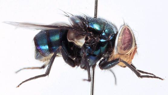
Identification of adult flies is seldom required for the diagnosis of myiasis, because the larval stages are those most apparent to livestock owners and veterinary personnel. However, a brief description follows:- The body is up to 10 mm long and has a metallic blue, bluish-purple or blue-green colour. The lower squamae are distinctly covered with fine hairs over their entire upper surface in OWSF and other Chrysomya species. Adults of OWSF can be distinguished from other Chrysomya found in cases of myiasis by the combination of black-brown to dark-orange-coloured anterior thoracic spiracles (rather than pale yellow, creamy, or white), with waxy-white, lower squama (rather than blackish-brown to dirty-grey) (Spradbery, 1991; Zumpt, 1965). In addition to the standard morphological techniques discussed above, more recent techniques for identification of screwworms and their geographical origins include cuticular hydrocarbon analysis (Brown et al, 1998), analysis of mitochondrial DNA (Hall et al, 2001; Litjens et al, 2001; Taylor et al, 1996), and use of random amplified polymorphic DNA polymerase chain reaction (RAPD-PCR) (Skoda et al, 2002).
Biology
The two screwworm flies, share almost identical biologies and both are true obligate parasites of mammals. Unlike most other species of blowflies, adult female screwworms do not lay their eggs on carrion. Instead, they lay them at the edges of wounds on living, injured mammals or at their body orifices. Virtually any wound is attractive, whether natural (from fighting, predators, thorns, disease, and/or tick and insect bites) or man-made (from shearing, branding, castrating, de-horning, docking, and/or ear-tagging). Commonly infested natural wounds are the navels of newborn animals and the vulval and perineal regions of their mothers, especially if traumatised. If eggs are deposited on mucous membranes, the larvae can invade undamaged natural body openings such as the nostrils and associated sinuses, the eye orbits, mouth, ears, and genitalia. The peak of oviposition activity occurs in the 2-3 hours prior to dusk (Spradbery, 1979).
Within 12–24 hours of the eggs being laid, larvae emerge and immediately begin to feed on the wound fluids and underlying tissues, burrowing gregariously head-downwards into the wound in a characteristic screwworm fashion. As they feed, tearing the tissue with their hook-like mouthparts, the wound is enlarged and deepened, resulting in extensive tissue destruction. Infested wounds often emit a characteristic odour, which can be the first indication that at least one animal in a group is infested. Although the odour is not always apparent to humans, it is obviously highly attractive to gravid females (Hall, 1995), which lay further batches of eggs so increasing the extent of the infestation. A severe infestation that is left untreated may result in the death of the host.
Screwworm larvae pass through three stages (or instars), separated by cuticular moults that facilitate rapid growth, and they reach maturity about 5– 7 days after egg hatch. They then stop feeding and leave the wound, falling to the ground into which they burrow and pupariate. The main exodus of larvae from the wound is thought to occur at night (Spradbery, 1983). The pupa develops within the puparium, a barrel-shaped protective structure formed by hardening and darkening of the cuticle of the mature larva. On completion of development, adult flies usually emerge from the puparium in the morning and work their way up to the soil surface, where they extend their wings for hardening prior to flight. Experimental data suggests that fly emergence is timed to occur around dawn each day (Spradbery, 1982). The duration of the underground puparial stage is temperature dependent and can last from a week to a month or more, depending on the season.
Males become sexually mature and able to mate within 24 hours, but females need to mature their ovaries over 6-7 days, and they only become responsive towards males and mate when about 3 days old. About 4 days after mating, female flies are ready to oviposit. They seek a suitable host and lay their eggs, all oriented in the same direction, like a tiled roof, firmly attached to each other and to the oviposition substrate. The numbers of eggs laid per batch vary depending on many factors (e.g. fly strain, disturbance during oviposition), but the average first batch has in the order of 175 eggs for OWSF (Spradbery, 1994). Following the first egg batch, further batches are laid at intervals of 3–4 days. In the wild females rarely survive long enough to lay more than 2 egg batches. Males and females live on average for 2–3 weeks during which time they feed at flowers, and the females also take in protein, e.g. from serous fluids at animal wounds.
Distribution
The distribution of OWSF is confined to the Old World, as the name suggests, throughout much of Africa (from Ethiopia and sub-Saharan countries to northern South Africa), the Gulf countries, the Indian subcontinent, and South-East Asia (from southern China [People’s Rep. of] through the Malay Peninsula and the Indonesian and Philippine islands to Papua New Guinea)(James, 1947; Zumpt, 1965; Sutherst et al, 1989; Spradbery, 1994). OWSF was reported from Hong Kong for the first time in 2000, infesting dogs, and a first human case was reported from there in 2003 (Ng et al., 2003). The situation in the Gulf area and surrounding regions is dynamic with recent reports confirmed from Iran and Iraq (Al-Izzi, 1999; Navidpour, 1996; Siddig, 2005). Epidemics of traumatic myiasis can follow introductions into such areas, especially where the livestock owners and veterinarians are unfamiliar with OWSF.
Primary Sources
- Hall, M.J.R. (1991) Screwworm flies as agents of wound myiasis. Pp. 8 17 in, World Animal Review, Special Issue "New World Screwworm: Response to an Emergency", October 1991, R.D.S. Branckaert (Ed.) 52 pp.[ view on-line version of this document ]
- Hall, M.J.R. (2004). New World screwworm (Cochliomyia hominivorax) and Old World screwworm (Chrysomya bezziana). Chapter 2.2.8., pp.370-379, in, Manual of Diagnostic Tests and Vaccines for Terrestrial Animals (mammals, birds and bees), Fifth Edition, Volume 1. World Organisation for Animal Health, Paris, France, 1178 pp.[ view on-line version of this document ]
References
- Al-Izzi M.A.J., Al-Taweel A.A. & Jassim F.A. (1999). Epidemiology and rearing of Old World screwworm, Chrysomya bezziana Villeneuve (Diptera; Calliphoridae) in Iraq. Iraqi J. Agricul. 4: 153–160.
- Brown W.V., Morton R., Lacey M.J., Spadbery J.P. & Mahon R.J. (1998). Identification of the geographical source of adults of the Old World screw-worm fly, Chrysomya bezziana Villeneuve (Diptera: Calliphoridae), by multivariate analysis of cuticular hydrocarbons. Comp. Biochem. Physiol. 119 B: 391-399.
- Hall, M.J.R. (1995). Trapping the flies that cause myiasis: their responses to host-stimuli. Ann. Trop. Med. Parasitol. 89: 333–357.
- Hall M.J.R., Edge W., Testa J., Adams Z.J.O. & Ready P.D. (2001). Old World screwworm fly, Chrysomya bezziana, occurs as two geographical races. Med. Vet. Entomol. 15: 393-402.
- James M.T. (1947). The Flies that Cause Myiasis in Man. United States Department of Agriculture Miscellaneous Publication No. 631, USDA, 175 pp.
- Laake E.W., Cushing E.C. & Parish H.E. (1936). Biology of the Primary Screwworm Fly, Cochliomyia americana, and a Comparison of its Stages with those of C. macellaria. United States Department of Agriculture, Technical Bulletin No. 500, USA, 24 pp.
- Litjens P., Lessinger A.C., De Azeredo-Espin A.M.L. DE (2001). Characterization of the screwworm flies Cochliomyia hominivorax and Cochliomyia macellaria by PCR-RFLP of mitochondrial DNA. Med. Vet. Entomol. 15: 183-188.
- Navidpour Sh., Hoghooghi-Rad N., Goodarzi H & Pooladgar A.R. (1996). Outbreak of Chrysomyia bezziana in Khoozestan province, Iran. Vet. Rec. 139: 217.
- Ng K.H.L., Yip, K.T., Choi C.H., Yeung K.H., Auyeung T.W., Tsang A.C.C., Chow L. & Que T.L. (2003). A case of oral myiasis due to Chrysomya bezziana. Hong Kong Med. J. 9: 454-456.
- Siddig, A, Al Jowary, S., Al-Izzi M., Hopkins, J., Hall, M.J.R. & Slingenbergh, J. (2005). The seasonality of Old World screwworm (Chrysomya bezziana) in the Mesopotamia valley in Iraq. Medical and Veterinary Entomology 19: 140-150.
- Skoda S.R., Pornkulwat S. & Foster J.E. (2002). Random amplified polymorphic DNA markers for discriminating Cochliomyia hominivorax from C. macellaria (Diptera: Calliphoridae). Bull. Entomol. Res. 92: 89-96.
- Spradbery, J.P. (1979). Daily oviposition activity and its adaptive significance in the screw-worm fly, Chrysomya bezzinana (Diptera: Calliphoridae). J. Aust. Ent. Soc. 18: 63-66.
- Spradbery, J.P. (1982). Diel pattern of adult emergence in the screw-worm fly Chrysomya bezziana (Diptera: Calliphoridae). J. Aust. Ent. Soc.21: 301-302.
- Spradbery, J.P. (1983). Diel larval exodus in the screw-worm fly Chrysomya bezziana (Villeneuve). J. Aust. Ent. Soc. 22: 261-262.
- Spradbery, J.P. (1991). A Manual for the Diagnosis of Screw-worm Fly. Commonwealth Scientific and Industrial Research Organization (CSIRO) Division of Entomology, Canberra, Australia, 64 pp.
- Spradbery, J.P. (1994). Screw-worm fly: a tale of two species. Agric. Zoo. Rev., 6, 1–62.
- Sutherst R.W., Spradbery J.P. & Maywald G.F. (1989). The potential geographical distribution of the Old World screwworm fly, Chrysomya bezziana. Med. Vet. Entomol. 3: 273–280.
- Taylor D.B., Szalanski A.L. & Peterson R.D. II (1996). Identification of screwworm species by polymerase chain reaction-restriction fragment length polymorphism. Med. Vet. Entomol. 10 63-70.
- Zumpt F. (1965). Myiasis in Man and Animals in the Old World. Butterworths, London,UK, 267 pp.


